All posts by Cliff Reid

Oncology Patient in Resus

This is the third hypothetical case discussion based on real cases submitted to the Resuscitology Team for analysis and debrief on the Resuscitology Course. Course participants are asked: “Please describe a resuscitation case that you have been involved in or witnessed that you feel (a) either had great learning points for other people on the course or (b) you feel didn’t go as well as it should and would appreciate the chance to analyse and discuss it.”

Case details are removed or altered to guarantee anonymity and confidentiality while preserving learning points. Cases are shared with the permission of the submitting clinician.

This case discussion was facilitated and summarised by Chris Nickson

A young adult female presents to the emergency department in septic shock. She has a diagnosis of metastatic cancer and is receiving chemotherapy. She is critically ill, with an altered mental state and is hypotensive.

She has no advance directive and the oncology consultant advises that “full resuscitation” should be performed. The ED team are not completely at ease with this, however, they continue to resuscitate the patient.

Eventually, the decision is made to intubate the patient, who has no externally apparent features of difficult anatomy. Two operators are unsuccessful with intubation attempts despite the use of video laryngoscopy. The patient is able to be oxygenated and ventilated until an anaesthetist is available to assist with the intubation.

Discussion points

  • Recognition and initial management of septic shock, with specific considerations in oncology patients
  • Should this patient be “for resuscitation”? How much is “too much”?
  • How can we clarify the best course of action when there is uncertainty or differences of opinion?
  • Why might attempts at intubation with video laryngoscopy fail, and how should we manage this?

Recognition and initial management of septic shock

Definition and recognition of sepsis is detailed in the LITFL CCC (https://lifeinthefastlane.com/ccc/sepsis-definitions/). Key elements to note:

  • Layperson’ definition works best! Sepsis has 3 key components:
      • Infection
      • Dysregulated host response
      • Organ dysfunction
  • Recognition
      • Suspected infection, and:
          • Two of hypotension, altered mental state, and tachypnea (but lacks sensitivity) = sepsis
          • Sepsis with refractory hypotension + lactate >2 mM = septic shock
      • Beware of sepsis mimics…
  • Rule of thumb: suspect sepsis in any patient with new organ dysfunction not otherwise explained

Initial management is detailed in the LITFL CCC (https://lifeinthefastlane.com/ccc/initial-sepsis-management/) under these headings:

  • resuscitation
  • early administration of appropriate antibiotics following blood cultures
  • early source control
  • judicious fluid resuscitation, avoiding excess fluids
  • noradrenaline for refractory hypotension (septic shock)
  • inotropes for septic cardiomyopathy
  • therapies for refractory hypotension
  • other experimental and rescue therapies
  • ongoing supportive care and monitoring

What specific considerations are there in the oncology patient?

  • Consider sepsis mimics (see: http://www.emdocs.net/mimics-of-sepsis/)
  • Haemophagocytic lymphohistiocytosis (HLH) (see: https://lifeinthefastlane.com/ccc/haemophagocytic-lymphohistiocytosis-hlh/)
  • Drug reactions – e.g. trial drugs, ATRA syndrome (APML)
  • Neoplastic manifestations – e.g. Addison’s, hyperthyroidism, vasculitis
  • Distributive shock – e.g. Anaphylaxis, Addison’s (other causes of shock/ hypotension)
  • Endocrine – e.g. Addison’s, hyperthyroidism, DKA
  • Aspiration
  • Hepatic failure
  • Toxicology – e.g. Salicylate poisoning, vasoplegic overdose (e.g. ACEI), hyperthermic toxidromes
  • Nutritional deficiency – e.g. thiamine deficiency
  • Gastrointestinal disorders – e.g. bowel obstruction, ileus, gut ischemia

Likely sources of sepsis

  • Pneumonia, urinary tract infections, wounds
  • Don’t forget:
      • Deep abscess/ soft tissue infections
      • Neuraxial infection
      • Endocarditis
      • Skin (always look at the back!)
      • Intra-abdominal sepsis
      • Lines, devices, and indwelling catheters

Differences in the manifestations/ recognition of sepsis occur as oncology patients, and immunosuppressed patients, may lack normal inflammatory response

  • Sepsis may be present with absence of:
      • Fever
      • Leukocytosis
  • On the other hand, fever and abnormal white count may occur due to the underlying malignancy or drugs

Effect of ICU/ critical illness on cancer

Sepsis may directly impact on tumour growth (Staudinger and Pene, 2014)

Many ICU drugs are thought to promote neoplastic growth:

  • Catecholamines
  • Sedatives
  • Blood transfusion
  • Hypoxia

Critical illness may:

  • Worsen immunosuppression
  • Worsen frailty

Should a young patient with metastatic cancer be “for resuscitation”? How much is “too much”?

Key considerations

  • How certain is the information we have?
  • What type of metastatic cancer and is the chemotherapy curative or palliative?
  • What is the advice of the consultants and “where are they coming from”?
  • What is the prognosis?
  • What are the patient’s values?

How certain is the information we have?

  • The general rule is “resuscitate before you prognosticate”
  • When the information need to guide decision making is uncertain, an approach of ongoing resuscitation while actively gathering further information (e.g. from clinical notes, family, specialist teams) is usually wise. Life sustaining measures are reversible, death isn’t.
  • As a team leader in this situation, it is useful to acknowledge the team’s discomfort, share your mental model, and check in on the team. For example, “This is a difficult situation. The patient may have a terminal illness, however, we do not know enough about them to be clear that stopping resuscitation is the right thing to do. I think we should continue resuscitation until we know more. Team, tell me what you think.”

What does ”metastatic cancer” mean? Is the chemotherapy curative or palliative?

  • Not all cancer, and not all metastasis is the same!
  • Therapies and prognoses are rapidly evolving – what we learnt in medical school may no longer be true! (duh)
  • Example: malignant melanoma; advanced cancers may be essentially curable if they respond to modern therapies

What is the advice of the consultants and “where are they coming from”?

  • We should remember to treat others with positive regard! (most people are doing their best, and they often do what do due to their circumstances, not because they are “bad”)
  • We are all subject to cognitive biases (e.g. sunk costs) and affective biases (e.g. guilt, emotional attachment, sterotyping); and base our advice on different backgrounds of expertise and experience
  • For instance (note these are generalisations):
    Click to to enlarge table in new tab

What are the patient values?

  • An understanding of patient values is best established when the patient is “well” and documented in an advance directive.
  • Appropriately documented advance directives are legally binding in Victoria.
  • If the patient is not competent, the “next of kin”/ family may act as surrogates to advise what they think the patient would want out of life and what is important to them
  • The surrogates do not choose medical therapies, that is the job of a clinician – we should avoid asking “what do you want us to do?”.
  • In time-critical emergencies surrogates may find it difficult to separate their own emotions/ needs from that of the patient; a useful question for surrogates is “if X was somehow able to be present in this discussion right now, what would X say?”
  • Sometimes we have no guidance on patient values and we just have to do what we think is right based on the information we have – if we stop, the patient is dead; if we don’t stop we can stop later when further information comes to light…
  • These issues are discussed further in the LITFL CCC page on “Families that want everything done”: https://lifeinthefastlane.com/ccc/patients-families-want-everything-done/

In most situations, the prognosis of the critical illness overrides the prognosis of the oncological diagnosis

  • For most oncology patients with solid tumours admitted to ICU, the 6-12 month mortality is similar to non-oncology patients ((De Jong & Bos, 2009). Caveats to consider:
      • Oncology patients are not a homogenous group
      • Data tends to be negatively biased by higher mortality in older studies
      • In the 1990s, oncology patients were considered very high risk (up to 90%) of mortality. At that time, immediate treatment limitations or even refusal of ICU admission for these patients were advocated. This pessimism may persist in people who trained in critical care around that time. (De Jong & Bos, 2009)
  • As a rule of thumb; the number and severity of organ impairments is the best guide to prognosis. Other indicators include:
      • Need of mechanical ventilation (especially for acute respiratory distress syndrome)
      • Vasopressor support (>4 hours) and therapies that have preceded ICU admission
      • The underlying neoplasm seems to have a little impact on the outcome
  • The most frequent reasons leading a cancer patient to ICU are:
      • Postoperative recovery
      • Respiratory failure
      • Infection and sepsis
  • A suggested approach (Biskup et al, 2017; Kiehl et al, 2018):
      • Patients with >6 months to live should have consideration of admission to ICU for trial of ICU therapy, with reassessment of progress at 3-10 days, then consideration of palliation if not improving
      • Patients with <6 months to live should have consideration of palliation if their illness would otherwise meet criteria for ICU admission
      • However, this approach should be tailored to the patient’s values (if attending a daughter’s wedding in 1 week is a dying patient’s life goal, then aggressive life-sustaining measures until that time may be acceptable to them)
  • Cardiopulmonary resuscitation (CPR)
      • Traditionally, CPR has been deemed inappropriate for most cancer patients due to poor survival to discharge (6% overall) (Sehatzadeh et al, 2014). However, these data are likely biased by older studies.
      • A recent retrospective study of 133 patients with cancer who required CPR found that 17% were alive at hospital discharge with good neurological recovery. (Champigneulle et al, 2016). However, improvement in CPR survival of oncology patients may be due to more selective performance of CPR through better use of consensus resuscitation plans.

Why might attempts at intubation with video laryngoscopy fail, and how should we manage this?

Key considerations

  • Failed airway algorithm
  • How to optimise intubation attempts: “STOPME” mnemonic
  • Reasons why video laryngoscopy can fail

Failed airway algorithm – “ensure first attempt is best possible attempt”, for example:

  • Plan A – Video laryngoscopy (VL) or direct laryngoscopy (DL) (up to 3 attempts)
  • Plan B – supraglottic airway device (SAD) / bag-valve-mask (BVM) (can switch back to Plan A if in “green zone” (able to effectively provide alveolar oxygenation) and STOPME is performed)
  • Plan C — FONA (front of neck access; i.e. emergency surgical airway)

How to optimise intubation attempts using the “STOPME” mnemonic:

  • Suction
  • Tone (administer neuromuscular blocker)
  • Operator (change to most expert intubator available)
  • Position (“ear-to-sternal notch” / “sipping the pint” position)
  • Manoeuvres (bimanual laryngoscopy, head lift, two handed lift, jaw thrust)
  • Equipment type and size: DL vs VL (using either a MacIntosh/ “standard geometry” or a Hyperangulated blade) or flexiscope.

Remember that DL commonly rescues VL, and vice versa! It makes sense to use a standard geometry VL blade that can also be used for DL, in most circumstances.

Specific reasons for failure of VL technique (see LITFL CCC for a description of video laryngoscopy: (https://lifeinthefastlane.com/ccc/video-laryngoscopy/)

  • Education, training, and familiarity with specific VL equipment and technique for use
  • Poor ergonomics
      • Best if intubator and team can see screen in front of intubator (allows “shared mental model”)
  • Poor view
      • Need to use “Look in mouth / at screen/ in mouth / at screen” approach – avoid piercing the soft palate or tonsilar bed with a bougie or stylet!
      • Hyperangulated VL can be used for Column 1 issues (if able to insert in mouth), and Column 3 issues (e.g. stiff neck), but can make view worse/ tube delivery more difficult if these are not present (see Keith Greenland’s Two Curve Theory and Three Column approach here: https://lifeinthefastlane.com/ccc/three-axis-alignment-versus-two-curve-theory/)
      • Need optimal patient positioning for laryngoscopy (e.g. ear-to-sternal notch positioning)
      • Remember to use appropriate manoeuvres to optimise laryngoscopy (e.g. bimanual laryngoscopy, head lift, lift epiglottis vs seating in vallecula, two hands on laryngoscopy handle for forceful lift)
      • Avoid contaminating VL camera or light source with blood, vomitus, or secretions – suction as you advance the VL tip (“stay high and dry”)
  • Failure of tube delivery
      • Use a stylet with same curvature of a hyperangulated VL blade; this is generally better than a bougie, which tends to lose curvature
      • Keep laryngeal inlet in top third of screen and to the left of centre; allow bougie/ tube delivery to be visualised and allows room to manourvre tip through the laryngeal inlet (“stay high and dry, not too close”)
      • Deliver tube using a “stop, pop, drop” approach; to avoid the stylet/ ETT tip abutting the anterior wall of the trachea following passage through the laryngeal inlet, rotate stylet to the right when advancing into the trachea

Potential reasons for failure in this case based on the limited vignette alone (using STEPS and GPAS approaches)

STEPS approach:

Self/ Team Leader

  • Stress – interpersonal conflict, moral distress, “HALT” factors (hungry, angry, lonely, tired)
  • Experience/ expertise with technique

Team

  • As per self
  • Ability to speak up

Environment

  • Equipment availability and familiarity
  • Equipment setup and positioning

Patient

  • Positioning
  • Difficult laryngoscopy – LEMON: tumors (mouth, compressing or obstructing airway), radiation

System

  • Availability of support and equipment
  • Lack of training
  • Culture (e.g. psychological safety and speaking up, silos)

GPAS approach:

Goal

  • Resuscitation of septic shock (should resuscitation be performed? Is it septic shock?)

Plan (Strategy)

  • Management of septic shock? (ABCs, culture, antibiotics, Rx haemodynamics, source control)

Actions (Tactics)

  • Consult with oncology team (and others?)
  • Airway assessment, timing and indications of intubation?

Skills

  • Effective communication skills? Right people?
  • Airway assessment skills?
  • VL skills?

References


Biskup E, Cai F, Vetter M, Marsch S. Oncological patients in the intensive care unit: prognosis, decision-making, therapies and end-of-life care. Swiss Med Wkly. 2017;147:w14481. [article]


Champigneulle B, Cariou A, Vincent F. Cardiopulmonary Resuscitation and Benefit to Patients With Metastatic Cancer. JAMA Intern Med. 2016;176(1):142. [article]


De jonge E, Bos MM. Patients with cancer on the ICU: the times they are changing. Crit Care. 2009;13(2):122. [article]


Kiehl MG, Beutel G, Böll B, et al. Consensus statement for cancer patients requiring intensive care support. Ann Hematol. 2018;97(7):1271-1282. [article]


Mack JW et al. End-of-Life Care Discussions Among Patients With Advanced Cancer. A Cohort Study. Ann Intern Med 2012;156:204-210. [article]


Nassar AP, Dettino ALA, Amendola CP, Dos santos RA, Forte DN, Caruso P. Oncologists’ and Intensivists’ Attitudes Toward the Care of Critically Ill Patients with Cancer. J Intensive Care Med. 2017;:885066617716105. [article]


Nickson CP. Sepsis Definitions and Diagnosis. LITFL Critical Care Compendium. Available at URL: https://lifeinthefastlane.com/ccc/sepsis-definitions/ [Accessed 14 December 2018]


Nickson CP. Patients and Families that “want everything done”. LITFL Critical Care Compendium. Available at URL: https://lifeinthefastlane.com/ccc/patients-families-want-everything-done/ [Accessed 14 December 2018]


Nickson CP. Initial sepsis management. LITFL Critical Care Compendium. Available at URL: https://lifeinthefastlane.com/ccc/initial-sepsis-management/ [Accessed 14 December 2018]


Nickson CP. Hemophagocytic lymphohistiocytosis (HLH). LITFL Critical Care Compendium. Available at URL: https://lifeinthefastlane.com/ccc/haemophagocytic-lymphohistiocytosis-hlh/ [Accessed 14 December 2018]


Sehatzadeh S. Cardiopulmonary Resuscitation in Patients With Terminal Illness: An Evidence-Based Analysis. Ont Health Technol Assess Ser. 2014;14(15):1-38. [article]


Staudinger T, Pène F. Current insights into severe sepsis in cancer patients. Rev Bras Ter Intensiva. 2014;26(4):335-8. [article]

Deterioration Post-MET Call

This is the second hypothetical case discussion based on real cases submitted to the Resuscitology Team for analysis and debrief on the Resuscitology Course. Course participants are asked: “Please describe a resuscitation case that you have been involved in or witnessed that you feel (a) either had great learning points for other people on the course or (b) you feel didn’t go as well as it should and would appreciate the chance to analyse and discuss it.”

Case details are removed or altered to guarantee anonymity and confidentiality while preserving learning points. Cases are shared with the permission of the submitting clinician.

This case discussion was facilitated and summarised by Cliff Reid

60 year old male with End Stage Renal Disease (ESRD). Admitted to ICU after a MET call for possible sepsis.


A formal echo showed severe LV systolic dysfunction, severe aortic stenosis, and RV dysfunction.


Three hours into the ICU admission he became peri-arrest. He was grey in colour, distressed and c/o central chest pain or pressure. He became hypotensive and was peri-arrest for about one hour, then arrested.


The patient’s young family was outside watching and crying. We had a lot of staff in the room and outside helping. Possibly too many, but it was useful to have a good line up for CPR. An airway was established early on in the code. And the person on the airway was watching the art line and ETCO2 for effective CPR and providing some coaching. It was chaotic, but we had good role allocation.


The cardiac arrest resuscitation lasted 30 minutes before being discontinued. He was mostly in a non-shockable rhythm.


There was a comment made afterwards, a concern by the person doing airway that he may have been aware/semi-conscious during part of the arrest.

Resuscitology Discussion Points

1. Cardiac arrest in ESRD patients.

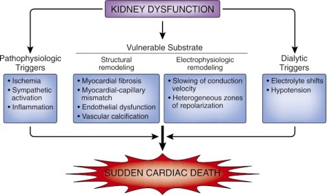
The prognosis from cardiac arrests is even worse in patients with kidney dysfunction in which survival probability decreases with a declining GFR. Among patients with ESRD who have a witnessed cardiac arrest at an outpatient dialysis facility, more than three-quarters are not discharged alive from the hospital. Structural and electrophysiologic remodeling of the heart, vascular calcification and fibrosis, autonomic dysregulation, and volume and electrolyte shifts are some of the underlying processes thought to explain the increased predisposition for sudden cardiac death in people with chronic kidney disease.(1)

Image from I.R. Whitman, H.I. Feldman, R. Deo. CKD and sudden cardiac death: epidemiology, mechanisms, and therapeutic approaches. J Am Soc Nephrol, 23 (2012), pp. 1929-1939

2. Establishing goals of care

This patient has significant co-morbidity and severe physiological derangement and is therefore likely to have a grim prognosis. It is key to establish the patient’s wishes and minimise unnecessary suffering or futile interventions. On the other hand, curable causes of deterioration such as sepsis or pulmonary embolism may be present and early aggressive resuscitation and investigation are likely to be appropriate for this chronologically relatively young patient.

Often a parallel approach is required: aggressive resuscitation at the bedside and a concurrent discussion about the patient’s wishes with the relatives. A useful question to pose to relatives, used by Chris Nickson (often more appropriate further along into an ICU stay) is: “If he could talk to us now, what would he say about this?

3. Likely causes of his arrest and their prevention / management

The underlying causes of haemodynamic deterioration can be rapidly differentiated into problems of relative or absolute hypovolaemia, pump dysfunction, or obstructive causes (pulmonary embolism, tamponade, tension pneumothorax)(2). Clinical assessment combined with point of care ultrasound (POCUS) allows immediate selection of fluids, vasopressors, inotropes, cardiac management, or surgical or decompressive procedures. An early blood gas can identify haemodynamically significant electrolyte derangement, in particular ionised hypocalcaemia.

In this patient’s case the presence of both severe aortic stenosis and right ventricular dysfunction contribute to significant haemodynamic management challenges. He may require emergent procedural intervention such as balloon aortic valvuloplasty and senior intensive care, cardiothoracic, and cardiology personnel would need to be mobilised. In a non-tertiary centre or middle-of-the-night scenario this could bring its own challenges and frustrations.

4. Team and communication factors

Following admission to ICU, he was peri-arrest for around one hour prior to arresting. The management of a crashing hypotensive patient like this requires a team approach and an appropriate degree of resuscitative momentum. It is critical that senior doctors and nurses are mobilised to the bedside and that the mental model is shared that immediate resuscitation is required. If this is not happening then appropriately assertive but non-judgmental / non-confrontational language should be used by the nursing or junior medical staff to highlight the urgency of the case. Examples are:

  • “I am very concerned about this patient who is deteriorating”
  • “I need your help immediately with the assessment of this severely shocked patient”
  • “Do you want me to start adrenaline or noradrenaline while you echo him to work out what’s going on?”
  • “I’ll call the registrar/fellow/consultant as it looks like we’re going to need lots of pairs of hands”

5. Management of the cardiac arrest on the ICU

The presence of invasive arterial monitoring allows chest compressions to be optimised in terms of location, rate and depth to titrate to an optimal diastolic blood pressure to support coronary perfusion. It allows early identification of ROSC and also titration of vasopressor doses to a target blood pressure rather than blindly administering 1mg doses of adrenaline (3). Continuous waveform capnography allows identification of tracheal tube placement and, like arterial blood pressure monitoring, can assist in optimisation of chest compressions, and identification of ROSC.

Crowd control is important. As additional team members arrive at different times but all wanting information regarding the case and their role, it can be helpful to allocate a ‘meet and greet’ person to update them and to protect the team leader from distraction. This ‘Resus Marshal’ can also police the number of people at the bedside and ensure non-contributors remain ‘behind the line’.

6. The ‘awake’ CPR patient

If CPR is sufficiently effective to perfuse the brain (its goal) then awareness and even full consciousness is possible. This has certainly been witnessed many times with mechanical CPR devices. While it is likely that staff may be more likely to remember this than the patient, the inevitable discomfort and distress can be mitigated with sedatives and analgesics. Ketamine provides both and would be the recommendation of most of the Resuscitology faculty. Fentanyl and/or midazolam may be considered as alternatives or in addition to ketamine. Some services have developed protocols for this situation(4).

7. Learning as a team from suboptimal cases

A full debrief is ideal but often hard to achieve. A ‘hot debrief’, conducted briefly  by the team immediately after the event, can identify learning points and also review what worked well, leading to action points to take forward. Once such method is the ‘STOP 5’ debrief developed in Edinburgh by Dr Craig Walker’s team. A version used in Sydney is shown here:

Modified STOP5 Hot Debrief Tool based on that developed by Drs Craig Walker & Robert Jack, Edinburgh Royal Infirmary

References

1. I.R. Whitman, H.I. Feldman, R. Deo. CKD and sudden cardiac death: epidemiology, mechanisms, and therapeutic approaches. J Am Soc Nephrol, 23 (2012), pp. 1929-1939

2. Desbiens NA. Simplifying the diagnosis and management of pulseless electrical activity in adults: a qualitative review. Crit Care Med 2008;36(2):391-6

3. Meaney PA, Bobrow BJ, Mancini ME, Christenson J, de Caen AR, Bhanji F, et al. Cardiopulmonary Resuscitation Quality: Improving Cardiac Resuscitation Outcomes Both Inside and Outside the Hospital: A Consensus Statement From the American Heart Association. Circulation. 2013 Jul 22;128(4):417–35

4. Rice DT, Nudell NG, Habrat DA, Smith JE, Ernest EV. CPR induced consciousness: It’s time for sedation protocols for this growing population. Resuscitation. 2016 Jun;103:e15–6

Unsurvivable Injury In The ED

This is the first of a regular posting of hypothetical cases based on real cases submitted to the Resuscitology Team for analysis and debrief on the Resuscitology Course. Course participants are asked: “Please describe a resuscitation case that you have been involved in or witnessed that you feel (a) either had great learning points for other people on the course or (b) you feel didn’t go as well as it should and would appreciate the chance to analyse and discuss it.”

Case details are removed or altered to guarantee anonymity and confidentiality while preserving learning points. Cases are shared with the permission of the submitting clinician.

This case discussion was facilitated and summarised by Chris Nickson

Male in his 20s, Motorcycle accident. No pre-arrival notification. Arrived in very busy ED (2 arrests ongoing including a paediatric arrest). Traumatic brain injury and cardiac arrest at scene then ROSC.

Patient suffered devastating head injury and severe chest trauma. Profoundly hypoxic, with SpO2 persistently in the 70s despite being intubated on 100% oxygen with bilateral intercostal catheters.

The CT showed severe subarachnoid haemorrhage and fractured skull and tonsillar herniation (coning). The patient’s (English speaking) family was overseas. It was a highly emotive situation with 3 cardiac arrests in ED within an hour.


Clinical Priorities in the Resus Room

The overall goals in this scenario include:

  • Management of the entire ED, ideally so that multiple critical patients can receive optimal treatment (the senior doctor in charge of the ED may be best being “hands off” and maintaining situational awareness of the whole department)
  • Optimally resuscitate the patient pending determination of whether palliation is appropriate as soon as possible. Key clinical priorities here are to:
    • Address hypoxia, in light of a severe traumatic brain injury
    • Expedite imaging to clarify diagnosis and prognosis
  • Support staff during and after an extremely difficult situation

Aggressive resuscitation of a patient who is suspected to have non-survivable injuries can cause individual cognitive dissonance and tension within the team. In general, it is important to “resuscitate before you prognosticate”. It may help to explicitly state and acknowledge this openly with the team, e.g. “Team, this is a difficult situation. The patient may have non-survivable injuries, we need to determine this as soon as possible. Until we know for sure, we will resuscitate him as best we can.”

Causes of hypoxia in major trauma

In an intubated patient always remember to check DOPES first:

  • Displaced endotracheal tube
  • Obstructed endotracheal tube
  • Patient factors
  • Equipment (e.g. remove from ventilator)
  • Stacked breaths (i.e. dynamic hyperinflation); Stomach (need NGT in small children as gastric distention can cause respiratory embarrassment); Synchrony (between patient and ventilator)


Patient factors in this case can be:

  • Exacerbation of underlying disease (eg asthma), or
  • Trauma-related


Causes of hypoxia resulting from major trauma include:

  • Airways
    • Obstruction, injury
  • Lung
    • Contusion, laceration, haemorrhage, pneumatocoeles
    • Aspiration, Foreign bodies
    • Acute Respiratory Distress Syndrome (ARDS)
  • Pleural
    • Pneumothorax (tension, open, closed), hemothorax, chylothorax
  • Vessels
    • Pulmonary embolism, fat embolism, pulmonary vascular injury
    • Traumatic shunt or exacerbation of existing shunt (e.g. ASD/PFO, which can cause platypnea-orthodeoxia syndrome)
  • Neuromuscular
    • CNS injury, spinal injury, phrenic nerve injury
  • Systemic
    • Severe hypercapnia (“CO2 filled lungs”) (e.g. due to brain impact apnoea prior to assisted ventilation)
    • metHb causing falsely low SpO2 and “anaemic hypoxia” (smoke)


In Traumatic Brain Injury (TBI), proposed mechanisms of hypoxia (apart from brain impact apnea) include:

  • Acute neurogenic pulmonary edema
  • Sympathetically induced pulmonary vasoconstriction leading to V/Q mismatch
  • Pulmonary shunting with changes in intracranial hypertension, e.g. passive opening of pulmonary arterio-venous shunts

Brain impact apnoea is a potential cause of death immediately after TBI. Such patients may have a normal, or near normal, CT brain and if adequately supported can make a full recovery. Without intervention, apnoea may lead to death.


Management of refractory hypoxaemia in a TBI patient

Observational studies consistently show that hypoxaemia is associated with worse outcomes including increased mortality following severe TBI. Evidence of harm from hyperoxia is less consistent. Current BTF guidelines do not specify a target SpO2 for severe TBI.

A step-wise approach to post-intubation hypoxaemia is described in LITFL’s Critical Care Compendium. Lung recruitment manoeuvres are controversial, and have primarily been studied as part of an open lung approach to mechanical ventilation in ARDS. They are worth considering as a rescue technique for refractory hypoxaemia with lung infiltrates as they can sometimes elicit a physiological response, even in the absence of ARDS (e.g. lung collapse), though it is uncertain if they improve patient outcomes. There are numerous other ways to improve oxygenation in ARDS, some of which could be considered in non-ARDS patients.

Unless there is obstructive lung disease (e.g. asthma, COPD), a protective lung ventilation strategy is generally appropriate for both ARDS and non-ARDS patients (e.g. patients with pulmonary contusions). For example, low tidal volumes (4-8 mL/kg PBW with plateau pressures <30 cmH20). However, this approach typically allows for permissive hypercapnia, which is not optimal for TBI patients as hypercapnia promotes cerebral vasodilatation and raised intracranial pressure (ICP) (Della Torre et al, 2017). High PEEP settings can be used to treat hypoxia in TBI patients, despite a raised ICP. However, excessive PEEP sometimes worsens oxygenation (e.g. overdistention in unilateral disease may increase shunt). In lungs with normal compliance, usually no more than 25% of the PEEP is transmitted to the central veins. In poorly compliant lungs (e.g. ARDS) very little PEEP is transmitted to the ICP. A small study found that PEEP up to 15 cmH20 has minimal changes in ICP and no change in CPP with normal lungs (McGuire et al, 1997). Furthermore, another small study showed that PEEP up to 15 cmH20 improves brain tissue oxygenation in severe TBI patients with ARDS (Nemar et al, 2017). A greater concern is the effect of PEEP on haemodynamics (Doblar et al, 1981). High PEEP – especially in hypovolaemic patients (e.g. haemorrhage) and patients with right heart failure – can decrease venous return, decreasing cardiac output and MAP. The decrease in MAP will decrease CPP and contribute to secondary insults in TBI. However, the decrease in MAP from PEEP can usually be prevented or corrected by appropriate use of vasopressors (e.g. noradrenaline) and fluid resuscitation (e.g. blood in a trauma patient). Overall, a PEEP of 15 cmH20 appears safe in TBI patients, and higher levels of PEEP could be considered in patients with refractory hypoxaemia as the harm from hypoxaemia may outweigh the potential harm from raised ICP and correctable effects on MAP.

In TBI, maintain cerebral perfusion pressure (CPP = MAP – ICP) in usual ways and decrease O2 consumption:

Head at 30 degrees (or greater if severely increased ICP) Avoid neck constrictions (e.g. loosen C-spine collar and ETT ties if needed)

  • Avoid hypoxia (e.g. target SpO2 >95%) with FiO2 and PEEP (up to 15cm H20, consider higher if needed)
  • Target PCO2 35-40 mmHg (avoid both hyperventilation and hypoventilation)
  • Assume ICP 20 mmHg, so need MAP 80-90 mmHg for CPP 60-70 mmHg
  • Noradrenaline to maintain MAP (+/- ongoing haemostatic resuscitation if needed)
  • Consider hypertonic saline or mannitol, if ICP >20 mmHg or evidence of coning
  • Sedation and paralysis
  • Maintain euthermia (T 36-37C)
  • Blood glucose 6-10 mmol/L


Resuscitation room palliation

The CEASE mnemonic has been proposed as a systematic approach to discontinuing resuscitation:

  • Clinical factors
  • Effectiveness of resuscitation
  • Ask others (but not family)
  • Stop resuscitation efforts
  • Explain to family

The decision to take the patient to the CT scan, even with suboptimal oxygen saturations, is appropriate.

  • CT brain confirms the severity of the TBI and impending coning
  • provides closure for team and family
  • If patient comfort is maintained, the harm to the patient is minimised, and the risk of incorrect diagnosis/ prognosis is avoided (e.g. atropine given pre-hospital for bradycardia causing “fixed dilated pupils”)

Attempts to optimise oxygenation should be time-specified and task-specified. If all reasonable options have been tried, and sufficient time allowed to check for improvement, then it is important not to “flail” but to proceed to the next step (CT brain to confirm diagnosis and guide ongoing management).

The mantra “resuscitate before you prognosticate” applies. In this situation, there may be tension in the team between ongoing attempts at aggressive resuscitation and the expectation of probable palliation. Aggressive therapy should continue until the appropriateness of palliation is confirmed. Furthermore, if the patient’s wish was to be an organ donor, ongoing organ support will be in the patient’s interest even after brain death.

Prognosis is always challenging, and should involve a neurosurgical opinion. We should avoid giving up hope purely on the basis of “bilaterally fixed dilated pupils”, because favourable outcomes are possible (Scotter et al, 2015) (especially if an extradural haemorrhage is present).

Palliation can be performed in the emergency department, but suitability is context-dependent (e.g. clinical suitability, how busy the ED is, available quiet space and meeting rooms, family dynamics, etc). Transfer to ICU for ongoing care and/or consideration of organ donation may be appropriate.

Breaking Bad News on the Telephone

The first rule of breaking bad news is: do not do it over the phone. However, in some situations – such as family being overseas – it is unavoidable.

Here is an approach:

  • Rehearse before making the call (e.g. with a social worker, or someone else skilled in difficult conversations)
  • Although this needs to be done in a timely fashion, delay the phone call until you are psychologically prepared if at all possible
  • Check the identity of the patient and the identity of the NOK, including contacts details
  • Introduce yourself clearly (Name, Role, Hospital)
  • Check that you are speaking to the right person and that he or she is an adult
  • Be direct and compassionate, use the “D word” – for example say “I’m sorry that I have to tell you the worst possible news. Your son, Mike, died in a car crash tonight.”
  • Check if they have support… if they don’t, offer to call someone for them
  • Provide follow up (e.g. social worker contact number)

Hot tip (from Vera Sistenich):

  • The person involved in an emotionally draining resuscitation doesn’t have to be the person who breaks bad news to the family.
  • A senior colleague who was not emotionally involved in the case may be better placed to have the discussion.

The VitalTalk app (and website) is very useful for helping to develop your communication skills about serious illness with patients and families.

Team Debrief and Counselling

After an arrest, make sure loose ends are TIED up:

  • Team check (is everyone OK?)
  • Ingest and imbibe (if possible, take a break to eat, drink, and recharge)
  • Equipment resupply (be ready the next emergency)
  • Debrief (soon after the event)

You might also want to conduct a “pause”. Taking a moment as a team to acknowledge the life of the person who has died and the efforts of the team in trying to save them.

“Team check” means checking in on how team members are coping. They may say they are fine, but look for signs of stress or distress. If present, affected team members may need to take a break or even go home early.

An approach to “hot debriefs” (immediately after the event), along with links to references on the topic, is described on INTENSIVE. “Closing the loop” after the “hot debrief” should include checking in on people affected by the event and providing access to counselling, following up a patient’s outcome, reporting a sentinel event, or instigating a guideline change.


References

Bartels J. About the Medical Pause. ThePause.me. Accessed 12 May 2018. Available at URL: https://thepause.me/2015/10/01/about-the-medical-pause/

Della Torre V, Badenes R, Corradi F, et al. Acute respiratory distress syndrome in traumatic brain injury: how do we manage it?. J Thorac Dis. 2017;9(12):5368-5381. Available at URL:  https://www.ncbi.nlm.nih.gov/pmc/articles/PMC5756968/

Doblar DD, Santiago TV, Kahn AU, Edelman NH. The effect of positive end-expiratory pressure ventilation (PEEP) on cerebral blood flow and cerebrospinal fluid pressure in goats. Anesthesiology. 1981;55(3):244-50. [pubmed]

Mcguire G, Crossley D, Richards J, Wong D. Effects of varying levels of positive end-expiratory pressure on intracranial pressure and cerebral perfusion pressure. Crit Care Med. 1997;25(6):1059-62. [pubmed]

Nemer SN, Caldeira JB, Santos RG, et al. Effects of positive end-expiratory pressure on brain tissue oxygen pressure of severe traumatic brain injury patients with acute respiratory distress syndrome: A pilot study. J Crit Care. 2015;30(6):1263-6. [pubmed]

Nickson CP. Brain Impact Apnoea. Critical Care Compendium, Lifeinthefastlane.com. Accessed12 May 2018. Available at URL: https://lifeinthefastlane.com/ccc/brain-impact-apnoea/

Nickson CP. Brain Impact Apnoea. Critical Care Compendium, Lifeinthefastlane.com. Accessed 12 May 2018. Available at URL: https://lifeinthefastlane.com/ccc/oxygen-saturation-targets-critical-care/

Nickson CP. Amazing and Awesome “Hot Debriefs” after Critical Incidents. INTENSIVEblog.com. Accessed 12 May 2018. Available at URL: http://intensiveblog.com/amazing-awesome-hot-debriefs-critical-incidents/

Nickson CP. Lung recruitment manoevres. Critical Care Compendium, Lifeinthefastlane.com. Accessed 12 May 2018. Available at URL: https://lifeinthefastlane.com/ccc/recruitment-manoeuvres-in-ards/

Nickson CP. Platypnea-Orthodeoxia Syndrome. Critical Care Compendium, Lifeinthefastlane.com. Accessed 12 May 2018. Available at URL: https://lifeinthefastlane.com/ccc/platypnea-orthodeoxia-syndrome/

Nickson CP. Post-intubation hypoxia. Critical Care Compendium, Lifeinthefastlane.com. Accessed 12 May 2018. Available at URL: https://lifeinthefastlane.com/ccc/post-intubation-hypoxia/

Nickson CP. Improving Oxygenation in ARDS. Critical Care Compendium, Lifeinthefastlane.com. Accessed 12 May 2018. Available at URL: https://lifeinthefastlane.com/ccc/improving-oxygenation-in-ards/

Nickson CP. Breaking Bad News to Relatives. Critical Care Compendium, Lifeinthefastlane.com. Accessed 12 May 2018. Available at URL: https://lifeinthefastlane.com/ccc/breaking-bad-news-relatives/

Nickson CP. Protective Lung Ventilation. Critical Care Compendium, Lifeinthefastlane.com. Accessed 12 May 2018. Available at URL: https://lifeinthefastlane.com/ccc/protective-lung-ventilation/

Scotter J, Hendrickson S, Marcus HJ, Wilson MH. Prognosis of patients with bilateral fixed dilated pupils secondary to traumatic extradural or subdural haematoma who undergo surgery: a systematic review and meta-analysis. Emerg Med J. 2015;32(8):654-9. [pubmed]

Torke AM, Bledsoe P, Wocial LD, Bosslet GT, Helft PR. CEASE: a guide for clinicians on how to stop resuscitation efforts. Ann Am Thorac Soc. 2015;12(3):440-5.  Available at URL:  https://www.atsjournals.org/doi/full/10.1513/AnnalsATS.201412-552PS